There are two types of ECMO. The VA ECMO is connected to both a vein and an artery and is used when there are problems with both the heart and lungs. The VV ECMO is connected to one or more veins, usually near the heart, and is used when the problem is only in the lungs.
USCF is also now using a smaller portable ECMO device that is light enough to be carried by one person and can be transported in an ambulance or helicopter, making it possible to provide ECMO relief in emergency cases.
When is ECMO used:
Being placed on ECMO requires a surgical procedure but it is usually done in a patient's room. The patient is sedated and given pain medication and an anti-coagulant to minimize blood clotting. A surgeon, assisted by an operating room team, inserts the ECMO catheters into either an artery or veins. An x-ray is then taken to ensure the tubes are in the right place. Usually a patient on the ECMO pump will also be on a ventilator, which helps the lungs to heal. While on ECMO, the patient will be monitored by specially trained nurses and respiratory therapists, as well as the surgeon and surgical team. Since you will be sedated and have a breathing tube in place, supplemental nutrition will be provided either intravenously or though a nasal-gastric tube. Nutrition is delivered either intravenously or though a nasal-gastric tube
While on ECMO, you may be given certain medications including: heparin to prevent blood clots; antibiotics to prevent infections; sedatives to minimize movement and improve sleep; diuretics to help the kidney get rid of fluids; electrolytes to maintain the proper balance of salts and sugars; and blood products to replace blood loss. Discontinuing ECMO requires a surgical procedure to remove the tubes. Multiple tests are usually done prior to the discontinuation of ECMO therapy to confirm that your heart and lungs are ready. Once the ECMO cannulas are removed, the vessels will need to be repaired. This can be done either at the bedside or in the operating room. The doctor will use small stitches to close the spot where the tubes were placed. You will be asleep and monitored for this process. Even though you are off the ECMO, you may still need to be on a ventilator.
ECMO does carry risks including:
ECMO was first used successfully in the USA in 1976 and was introduced into this country in 1989. It was first set
up at GOSH in 1992 and to date we have
supported over 850 babies and children. GOSH is part of a national service
providing cardiorespiratory ECMO support for babies and children in England and Wales. The other centres are Glenfield
Hospital in Leicester, Birmingham Children's Hospital, Alder Hey Children's Hospital and the Freeman Hospital in
Newcastle.
ECMO is used for babies and children with severe heart (cardiac) or lung (respiratory) failure.
In children with very severe lung disease which is not responding to the usual treatment of mechanical ventilation (breathing machine), medicines and extra oxygen, ECMO can take over the function of the child’s lungs, allowing them time to rest and heal.
In children with very poor cardiac function, ECMO can take over the work of the child’s heart. This provides time for the heart to rest and recover, while maintaining a good blood supply to the brain and other organs in the body. Cardiac ECMO may be needed after open heart surgery, when the heart may be swollen, unable to maintain a high enough blood pressure or have an irregular rhythm. It may also be needed due to an infection affecting the heart muscle (myocarditis) or heart muscle failure (myopathy) where the heart cannot pump blood around the body effectively.
Please note that ECMO can only help children whose lung and/or heart disease is reversible within about three weeks.
ECMO may be started in the operating theatre immediately after surgery or on one of the intensive care units. If you
child is going onto ECMO after a heart
operation, the surgeon will usually insert the cannulae (tubes) during the
operation directly into the heart through the chest.
If ECMO is started in the intensive care unit, the cannulae connecting the patient to the ECMO circuit are placed directly into the blood vessels on the side of the neck. This short operation takes place at the bedside in the intensive care unit and you will be asked to leave during the procedure. A light anesthetic is given so your child does not feel any pain and a tube (cannula) is placed into a large vein in the side of the neck. A second tube is often required, which is inserted into an artery also on the side of the neck.
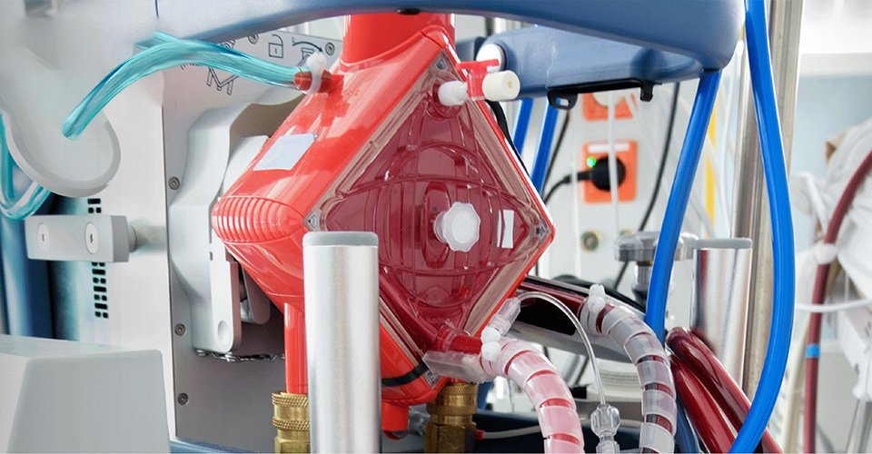
Once in place, the cannulae are then connected to the ECMO circuit and machine. Dark blood (containing little oxygen) drains from your child through the tube in the vein and is pumped through the membrane oxygenator (an artificial lung) where carbon dioxide is removed and oxygen added. The blood is then re-warmed and returned to the body. This process goes on continuously while your child is on ECMO. They will also stay on a ventilator but on very gentle settings which allow the lungs to rest.
There are two types of ECMO, VA (veno-arterial) and VV (veno-venous):
Two cannulae (tubes) are placed into your child’s blood vessels, one into a vein and the other into an artery, usually on the side of the neck. Dark blood (containing little oxygen) is drained continuously into the ECMO circuit from the tube in the vein while the now oxygen-rich blood from the ECMO circuit is returned to the body through the tube in the artery.
This type of ECMO provides support for both the heart and the lungs and so can be used for children requiring either cardiac or respiratory ECMO support. For the small number of children who require ECMO after open heart surgery, the cannulae (tubes) may be inserted directly into the heart through the chest during the heart operation rather than into the neck vessels.
A single catheter is placed into a vein, usually in the side of the neck. Blood is drained from this catheter into the ECMO circuit at the same time as oxygenated blood is returned through the same catheter from the ECMO circuit to the child. VV ECMO provides lung support only and does not support the heart.
A few children who start with VV ECMO will need to be changed over to VA ECMO if their heart also starts to need support.
Ecmo Healthcare